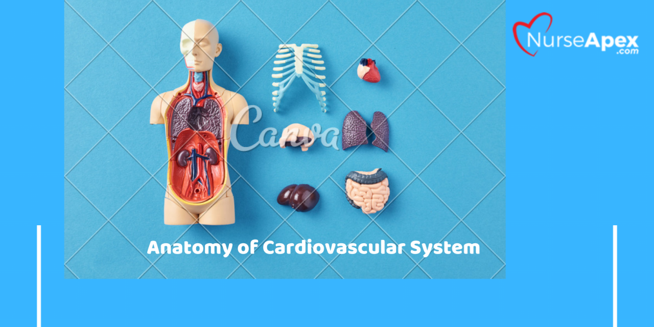
ECG for Healthcare Professionals, Chapter 2 The Cardiovascular System Posted by NurseApex | Jul 28, 2022 | General, Nursing Guide, Nursing Students, Study Notes | 0 | Score 80%Score 80% Action Potential The change in the electrical potential of the heart muscle when it is stimulated; depolarization followed by repolarization. Aorta The largest artery of the body; transport blood from the left ventricle of the heart to the entire body. Aortic Semilunar Valve Valve located in the aorta that prevents the backflow of blood into the left ventricle. Atrioventricular (AV) Node Delays the electrical impulse to allow the atria to complete their contraction. Atrium (Pl. Atria) Top two chambers of the heart. Automaticity The ability of the heart to initiate an electrical impulse without being stimulated by another or independent source. Bundle Branches Left and right branches pf the bundle of His that conduct impulse down either side of the interventricular septum, to the left and right ventricles. Bundle of His (AV Bundle) Located next to the AV node; provides the transfer of the electrical impulse from the atria to the ventricles. Cardiac Cycle The contraction and relaxation of the heart Complexes Atrial or ventricular contraction as they appear on the ECG; complete ECG waveforms. Conductivity The ability of the heart cells to receive and transmit an electrical impulse. Contractility The ability of the heart muscle cells to shorten in response to an electrical stimulus. Coronary Circulation The circulation of the blood to and from the heart. Deoxygenated Blood Blood that has little or minimal oxygen (oxygen-poor blood) Depolatization The electrical activation of the cells of the heart that initiates contraction of the heart muscle. Diastole The phase of the cardiac cycle when the heart is expanding and refilling; also known as the relaxation phase. Excitability The ability of the heart muscle cells to respond to an impulse or stimulus. Interval The period of time between two activities within the heart. Interventricular Septum A partition or wall (septum) that divides the right and left ventricles. Ischemia Lack of blood supply entrance into a body cavity, tissue, or blood vessel. Left Atrium The left upper chamber of the heart, which receives blood from the lungs. Left Ventricle The left lower chamber of the heart, which pumps oxygenated blood through the body, it is the biggest and strongest chamber, known as the workhorse of the heart. Mitral (Bicuspid) Valve Valve with two cups or leaflets located between the left atrium and left ventricle; it prevents backflow of blood into the left atrium. Myocardial Pertaining to the heart (cardio) muscle (myo) Oxygenated Blood Blood having oxygen (oxygen-rich blood) Pericardium A two-layered sac of tissue enclosing the heart. Polaraztion The stare of cellular rest in which the inside is negatively charge and the outside is positively charged. Pulmonary Artery Large artery that transports deoxygenated blood from the right ventricle to the lungs. This is the only artery in the body that carries deoxygenated blood. Pulmonary Circulation The transportation of blood to and from the lungs; blood is oxygenated in the lungs during pulmonary circulation. Pulmanary Semilaunar Valve A valve found in the pulmonary artery that prevents backflow of the blood back into the right ventricle during pulmonary circulation. Pulmoanary Vein Transports oxygenated blood back into the left atrium of the heart. Purkinje Fibers The fibers within the heart that distribute electrical impulses from cell to cell throughout the ventricles. Purkinje Network Spreads the electrical impulse throughout the ventricles by means of the Purlkinje fiber. Repolarization When heart muscle cells return to their resting electrical state and the heart muscle relaxes. Right Atrium The right upper chamber of the heart, which receives blood from the body. Right Ventricle The right lower chamber of the heart, which pumps blood to the lungs. Segment A portion or part of the electrical tracing produced by the heart. Semilunar Valve A valve with half-moon-shaped cusps that open and close, allowing blood to travel only one way; located in the pulmonary artery and the aorta. Sinoatrial (SA) Node An area of specialized cells in the upper right atrium that initiates the heartbeat. Systemic Circulation The pathways for pumping blood throughout the body and back to the heart. Systole The contraction phase of the cardiac cycle, during which the heart is pumping blood out of the body. Tricuspid Valve Valve located between the right atrium and right ventricle; it prevents backflow of blood into the right atrium. Vena Cava (Pl. Venae Cave) Largest vein in the body, which provides a pathway for deoxygenated blood to return to the heart; its upper portion, the superior vena cava, transports blood from the head, arms, and upper body; and its lower portion, the inferior vena cava, transports blood from the lower body legs. Facebook Google+ Like 0 Twitter Review 80% 80%


